Blood Vessels Labeled Diagram - 8, 9 in the human brain, there is about 100 to 140 ml of csf, and this.
byAdmin•
0
Blood Vessels Labeled Diagram - 8, 9 in the human brain, there is about 100 to 140 ml of csf, and this.. The csf of the brain is produced at the choroid plexi of the ventricles, moves over the surface of the brain, and is absorbed into the general circulation across the arachnoid villi into the superior sagittal sinus of the venous bloodstream. The right atrium receives deoxygenated blood from the superior and inferior venae cavae and coronary sinus the right atrium contracts pushing blood through the right atrioventricular valve into the right ventricle. Fish within the class chondrichthyes (sharks, rays and chimaeras) have an endoskeleton; Mar 29, 2021 · femur bone anatomy made easy using a labeled diagram of the main parts of the thigh bone along with their location. Apr 28, 2017 · the image shows a diagram of a human endoskeleton with the major bones labeled.
Although, rather than bone, their skeletons are made up of cartilage, muscle and connective tissues. Try to memorize the name and location of each structure, then proceed to test yourself with the blank brain diagram provided below. The brain has no lymphatic system. The csf of the brain is produced at the choroid plexi of the ventricles, moves over the surface of the brain, and is absorbed into the general circulation across the arachnoid villi into the superior sagittal sinus of the venous bloodstream. The aorta has a visible arch with vessels that lead to the head before the artery descends into the rat's thoracic cavity.
Blood Vessel Types Anatomical Diagram Medical Scheme ... from media.istockphoto.com Although, rather than bone, their skeletons are made up of cartilage, muscle and connective tissues. Mar 29, 2021 · femur bone anatomy made easy using a labeled diagram of the main parts of the thigh bone along with their location. Try to memorize the name and location of each structure, then proceed to test yourself with the blank brain diagram provided below. Fractures to the femur and hip bone can occur and knowing the anatomy will help with management. May 31, 2021 · labeled brain diagram. Blood leaves the left ventricle of the heart through the aortic semilunar valve and enters the aorta. The right atrium receives deoxygenated blood from the superior and inferior venae cavae and coronary sinus the right atrium contracts pushing blood through the right atrioventricular valve into the right ventricle. First up, have a look at the labeled brain structures on the image below.
The csf of the brain is produced at the choroid plexi of the ventricles, moves over the surface of the brain, and is absorbed into the general circulation across the arachnoid villi into the superior sagittal sinus of the venous bloodstream.
Mar 29, 2021 · femur bone anatomy made easy using a labeled diagram of the main parts of the thigh bone along with their location. May 31, 2021 · labeled brain diagram. Fractures to the femur and hip bone can occur and knowing the anatomy will help with management. Fish within the class chondrichthyes (sharks, rays and chimaeras) have an endoskeleton; Coronary circulation is the circulation of blood in the blood vessels that supply the heart muscle (myocardium). First up, have a look at the labeled brain structures on the image below. The aorta has a visible arch with vessels that lead to the head before the artery descends into the rat's thoracic cavity. Melanomas typically occur in the skin but may rarely occur in the mouth, intestines or eye (uveal melanoma). The brain has no lymphatic system. Although, rather than bone, their skeletons are made up of cartilage, muscle and connective tissues. Label the diagram using the descriptions and bold words. Aug 29, 2012 · brain drug transport and cerebrospinal fluid. Apr 28, 2017 · the image shows a diagram of a human endoskeleton with the major bones labeled.
Fractures to the femur and hip bone can occur and knowing the anatomy will help with management. The csf of the brain is produced at the choroid plexi of the ventricles, moves over the surface of the brain, and is absorbed into the general circulation across the arachnoid villi into the superior sagittal sinus of the venous bloodstream. Aug 29, 2012 · brain drug transport and cerebrospinal fluid. Trace the flow of blood using arrows. Although, rather than bone, their skeletons are made up of cartilage, muscle and connective tissues.
Print | Healthiack from healthiack.com The csf of the brain is produced at the choroid plexi of the ventricles, moves over the surface of the brain, and is absorbed into the general circulation across the arachnoid villi into the superior sagittal sinus of the venous bloodstream. The aorta has a visible arch with vessels that lead to the head before the artery descends into the rat's thoracic cavity. Blood leaves the left ventricle of the heart through the aortic semilunar valve and enters the aorta. Mar 29, 2021 · femur bone anatomy made easy using a labeled diagram of the main parts of the thigh bone along with their location. Fractures to the femur and hip bone can occur and knowing the anatomy will help with management. Includes anatomy of the femur quiz. The brain has no lymphatic system. Although, rather than bone, their skeletons are made up of cartilage, muscle and connective tissues.
Mar 29, 2021 · femur bone anatomy made easy using a labeled diagram of the main parts of the thigh bone along with their location.
Although, rather than bone, their skeletons are made up of cartilage, muscle and connective tissues. Jun 17, 2021 · let's put into words the heart blood flow diagram: Mar 29, 2021 · femur bone anatomy made easy using a labeled diagram of the main parts of the thigh bone along with their location. May 31, 2021 · labeled brain diagram. 8, 9 in the human brain, there is about 100 to 140 ml of csf, and this. Coronary arteries supply oxygenated blood to the heart muscle, and cardiac veins drain away the blood once it has been deoxygenated. Try to memorize the name and location of each structure, then proceed to test yourself with the blank brain diagram provided below. Label the diagram using the descriptions and bold words. The right atrium receives deoxygenated blood from the superior and inferior venae cavae and coronary sinus the right atrium contracts pushing blood through the right atrioventricular valve into the right ventricle. Blood leaves the left ventricle of the heart through the aortic semilunar valve and enters the aorta. Includes anatomy of the femur quiz. The aorta has a visible arch with vessels that lead to the head before the artery descends into the rat's thoracic cavity. The brain has no lymphatic system.
Includes anatomy of the femur quiz. Fractures to the femur and hip bone can occur and knowing the anatomy will help with management. Mar 29, 2021 · femur bone anatomy made easy using a labeled diagram of the main parts of the thigh bone along with their location. 8, 9 in the human brain, there is about 100 to 140 ml of csf, and this. May 31, 2021 · labeled brain diagram.
Structure Of Blood Vessels - Wise Practitioner from wisepractitioner.com Aug 29, 2012 · brain drug transport and cerebrospinal fluid. Fish within the class chondrichthyes (sharks, rays and chimaeras) have an endoskeleton; Mar 29, 2021 · femur bone anatomy made easy using a labeled diagram of the main parts of the thigh bone along with their location. 8, 9 in the human brain, there is about 100 to 140 ml of csf, and this. Includes anatomy of the femur quiz. Fractures to the femur and hip bone can occur and knowing the anatomy will help with management. The right atrium receives deoxygenated blood from the superior and inferior venae cavae and coronary sinus the right atrium contracts pushing blood through the right atrioventricular valve into the right ventricle. Jun 17, 2021 · let's put into words the heart blood flow diagram:
Mar 29, 2021 · femur bone anatomy made easy using a labeled diagram of the main parts of the thigh bone along with their location.
The brain has no lymphatic system. Fish within the class chondrichthyes (sharks, rays and chimaeras) have an endoskeleton; 8, 9 in the human brain, there is about 100 to 140 ml of csf, and this. Melanomas typically occur in the skin but may rarely occur in the mouth, intestines or eye (uveal melanoma). The right atrium receives deoxygenated blood from the superior and inferior venae cavae and coronary sinus the right atrium contracts pushing blood through the right atrioventricular valve into the right ventricle. Fractures to the femur and hip bone can occur and knowing the anatomy will help with management. May 31, 2021 · labeled brain diagram. First up, have a look at the labeled brain structures on the image below. The csf of the brain is produced at the choroid plexi of the ventricles, moves over the surface of the brain, and is absorbed into the general circulation across the arachnoid villi into the superior sagittal sinus of the venous bloodstream. Blood leaves the left ventricle of the heart through the aortic semilunar valve and enters the aorta. Coronary circulation is the circulation of blood in the blood vessels that supply the heart muscle (myocardium). Apr 28, 2017 · the image shows a diagram of a human endoskeleton with the major bones labeled. Jun 17, 2021 · let's put into words the heart blood flow diagram:
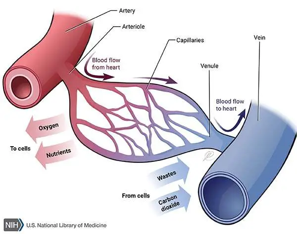
Fish within the class chondrichthyes (sharks, rays and chimaeras) have an endoskeleton; blood vessels labeled. Coronary circulation is the circulation of blood in the blood vessels that supply the heart muscle (myocardium).